-
13. Surface Anatomy of the Lower Extremity
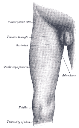
FIG. 1238 - Front and medial aspect of right thigh. Skin.—The skin of the thigh, especially in the hollow of the groin and on the medial side, is thin, smooth and elastic, and contains few hairs except on the neighborhood of the pubis. Laterally it is thicker and the hairs are more numerous. The junction of the skin of the thigh with that on the front of the abdomen is marked by a well-defined furrow which indicates the site of the inguinal ligament; the furrow presents a general convexity downward, but its medial half, which is the better marked, is nearly straight. The skin over the buttock is fairly thick and is characterized by its low sensibility and slight vascularity; as a rule it is destitute of conspicuous hairs except toward the post-anal furrow, where in some males they are abundantly developed. An almost transverse fold—the gluteal fold—crosses the lower part of the buttock; it practically bisects the lower margin of the Glutæus maximus and is most evident during extension of the hip-joint. The skin over the front of the knee is covered by thickened epidermis; it is loose and thrown into transverse wrinkles when the leg is extended. The skin of the leg is thin, especially on the medial side, and is covered with numerous large hairs. On the dorsum of the foot the skin is thin, loosely connected to subjacent parts, and contains few hairs, on the plantar surface, and especially over the heel, the epidermis is of great thickness, and here, as in the palm of the hand, there are neither hairs nor sebaceous glands. Bones.—The hip bones are largely covered with muscles, so that only at a few points do they approach the surface. In front the anterior superior iliac spine is easily recognized, and in thin subjects stands out as a prominence at the lateral end of the fold of the groin; in fat subjects its position is indicated by an oblique depression, at the bottom of which the bony process can be felt. Proceeding upward and backward from this process the sinuously curved iliac crest can be traced to the posterior superior iliac spine, the site of which is indicated by a slight depression; on the outer lip of the crest, about 5 cm. behind the anterior superior spine, is the prominent iliac tubercle. In thin subjects the pubic tubercle is very apparent, but in the obese it is obscured by the pubic fat; it can, however, be detected by following up the tendon of origin of Adductor longus. Another part of the bony pelvis which is accessible to touch is the ischial tuberosity, situated beneath the Glutæus maximus, and, when the hip is flexed, easily felt, as it is then uncovered by muscle. The femur is enveloped by muscles, so that in fairly muscular subjects the only accessible parts are the lateral surface of the greater trochanter and the lower expanded end of the bone. The site of the greater trochanter is generally indicated by a depression, owing to the thickness of the Glutæi medius and minimus which project above it; when, however, the thigh is flexed, and especially if it be crossed over the opposite one, the trochanter produces a blunt eminence on the surface. The lateral condyle is more easily felt than the medial; both epicondyles can be readily identified, and at the upper part of the medial condyle the sharp adductor tubercle can be recognized without difficulty. When the knee is flexed a portion of the patellar surface is uncovered and is palpable. The anterior surface of the patella is subcutaneous. When the knee is extended the medial border of the bone is a little more prominent than the lateral, and if the Quadriceps femoris be relaxed the bone can be moved from side to side. When the joint is flexed the patella recedes into the hollow between the condyles of the femur and the upper end of the tibia, and becomes firmly applied to the femur. A considerable portion of the tibia is subcutaneous. At the upper end the condyles can be felt just below the knee; the medial condyle is broad and smooth, and merges into the subcutaneous surface of the body below; the lateral is narrower and more prominent, and on it, about midway between the apex of the patella and the head of the fibula, is the tubercle for the attachment of the iliotibial band. In front of the upper end of the bone, between the condyles, is an oval eminence, the tuberosity, which is continuous below with the anterior crest of the bone. This crest can be identified in the upper two-thirds of its extent as a flexuous ridge, but in the lower third it disappears and the bone is concealed by the tendons of the muscles on the front of the leg. Medial to the anterior crest is the broad surface, slightly encroached on by muscles in front and behind. The medial malleolus forms a broad prominence, situated at a higher level and somewhat farther forward than the lateral malleolus; it overhangs the medial border of the arch of the foot; its anterior border is nearly straight, its posterior presents a sharp edge which forms the medial margin of the groove for the tendon of Tibialis posterior. The only subcutaneous parts of the fibula are the head, the lower part of the body, and the lateral malleolus. The head lies behind and lateral to the lateral condyle of the tibia, and presents as a small prominent pyramidal eminence slightly above the level of the tibial tuberosity; its position can be readily located by following downward the tendon of Biceps femoris. The lateral malleolus is a narrow elongated prominence, from which the lower third or half of the lateral surface of the body of the bone can be traced upward. On the dorsum of the tarsus the individual bones cannot be distinguished, with the exception of the head of the talus, which forms a rounded projection in front of the ankle-joint when the foot is forcibly extended. The whole dorsal surface of the foot has a smooth convex outline, the summit of which is the ridge formed by the head of the talus, the navicular, the second cuneiform, and the second metatarsal bone; from this it inclines gradually lateralward, and rapidly medialward. On the medial side of the foot the medial process of the tuberosity of the calcaneus and the ridge separating the posterior from the medial surface of the bone are distinguishable; in front of this, and below the medial malleolus, is the sustentaculum tali. The tuberosity of the navicular is palpable about 2.5 to 3 cm. in front of the medial malleolus. Farther forward, the ridge formed by the base of the first metatarsal bone can be obscurely felt, and from this the body of the bone can be traced to the expanded head; beneath the base of the first phalanx is the medial sesamoid bone. On the lateral side of the foot the most posterior bony point is the lateral process of the tuberosity of the calcaneus, with the ridge separating the posterior from the lateral surface of the bone. In front of this the greater part of the lateral surface of the calcaneus is subcutaneous; on it, below and in front of the lateral malleolus, the trochlear process, when present, can be felt. Farther forward the base of the fifth metatarsal bone is prominent, and from it the body and expanded head can be traced. As in the case of the metacarpals, the dorsal surfaces of the metatarsal bones are easily defined, although their heads do not form prominences; the plantar surfaces are obscured by muscles. The phalanges in their whole extent are readily palpable. Articulations.—The hip-joint is deeply seated and cannot be palpated. The interval between the tibia and femur can always be easily felt; if the knee-joint be extended this interval is on a higher level than the apex of the patella, but if the joint be slightly flexed it is directly behind the apex. When the knee is semiflexed, the medial borders of the patella and of the medial condyle of the femur, and the upper border of the medial condyle of the tibia, bound a triangular depressed area which indicates the position of the joint. The ankle-joint can be felt on either side of the Extensor tendons, and during extension of the joint the superior articular surface of the talus presents below the anterior border of the lower end of the tibia. Muscles.—Of the muscles of the thigh, those of the anterior femoral region (Fig. 1238) contribute largely to surface form. The Tensor fasciæ latæ produces a broad elevation immediately below the anterior part of the iliac crest and behind the anterior superior iliac spine; from its lower border a groove caused by the iliotibial band extends downward to the lateral side of the knee-joint. The upper portion of Sartorius constitutes the lateral boundary of the femoral triangle, and, when the muscle is in action, forms a prominent oblique ridge which is continued below into a flattened plane and then gradually merges into a general fulness on the medial side of the knee-joint. When the Sartorius is not in action, a depression exists between the Quadriceps femoris and the Adductors, and extends obliquely downward and medialward from the apex of the femoral triangle to the side of the knee. In the angle formed by the divergence of Sartorius and Tensor fasciæ lataæ, just below the anterior superior iliac spine, the Rectus femoris appears, and in a muscular subject its borders can be clearly defined when the muscle is in action. The Vastus lateralis forms a long flattened plane traversed by the groove of the iliotibial band. The Vastus medialis gives rise to a considerable prominence on the medial side of the lower half of the thigh; this prominence increases toward the knee and ends somewhat abruptly with a full curved outline. The Vastus intermedius is completely hidden. The Adductores cannot be differentiated from one another, with the exception of the upper tendon of Adductor longus and the lower tendon of Adductor magnus. When the Adductor longus is in action its upper tendon stands out as a prominent ridge running obliquely downward and lateralward from the neighborhood of the public tubercle, and forming the medial border of the femoral triangle. The lower tendon of Adductor magnus can be distinctly felt as a short ridge extending downward between the Sartorius and Vastus medialis to the adductor tubercle. The adductores fill in the triangular space at the upper part of the thigh, between the femur and the pelvis, and to them is due the contour of the medial border of the thigh, the Gracilis contributing largely to the smoothness of the outline. 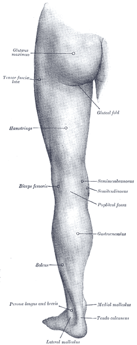
FIG. 1239 - Back of left lower extremity. The Glutæus maximus (Fig. 1239) forms the full rounded outline of the buttock; it is more prominent behind, compressed in front, and ends at its tendinous insertion in a depression immediately behind the greater trochanter; its lower border crosses the gluteal fold obliquely downward and lateralward. The upper is part of Glutæus medius visible, but its lower part with Glutæus minimus and the external rotators are completely hidden. From beneath the lower margin of Glutæus maximus the hamstrings appear; at first they are narrow and not well-defined, but as they descend they become more prominent and eventually divide into two well-marked ridges formed by their tendons; these constitute the upper boundaries of the popliteal fossa. The tendon of Biceps femoris is a thick cord running to the head of the fibula; the tendons of the Semimembranosus and Semitendinosus as they run medialward to the tibia are separated by a slight furrow; the Semitendinosus is the more medial, and can be felt in certain positions of the limb as a sharp cord, while the Semimembranosus is thick and rounded. The Gracilis is situated a little in front of them. The Tibialis anterior (Fig. 1240) presents a fusiform enlargement at the lateral side of the tibia and projects beyond the anterior crest of the bone; its tendon can be traced on the front of the tibia and ankle-joint and thence along the medial side of the foot to the base of the first metatarsal bone. The fleshy fibers of Peronæus longus are strongly marked at the upper part of the lateral side of the leg; it is separated by furrows from Extensor digitorum longus in front and Soleus behind. Below, the fleshy fibers end abruptly in a tendon which overlaps the more flattened elevation of Peronæus brevis; below the lateral malleolus the tendon of Peronæus brevis is the more marked. On the dorsum of the foot (Fig. 1241) the tendons emerging from beneath the transverse and cruciate crural ligaments spread out and can be distinguished as follows: the most medial and largest is Tibialis anterior, the next is Extensor hallucis proprius, then Extensor digitorum longus dividing into four tendons, to the second, third, fourth, and fifth toes, and lastly Peronæus tertius. The Extensor digitorum brevis produces a rounded outline on the dorsum of the foot and a fulness in front of the lateral malleolus. The Interossei dorsales bulge between the metatarsal bones. 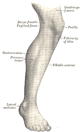
FIG. 1240 - Lateral aspect of right leg. At the back of the knee is the popliteal fossa, bounded above by the tendons of the hamstrings and below by the Gastrocnemius. Below this fossa is the prominent fleshy mass of the calf of the leg produced by Gastrocnemius and Soleus (Fig. 1239). When these muscles are in action the borders of Gastrocnemius form two well-defined curved lines which converge to the tendocalcaneus; the medial border is the more prominent. At the same time the edges of Soleus can be seen forming, on either side of Gastrocnemius, curved eminences, of which the lateral is the longer. The fleshy mass of the calf ends somewhat abruptly in the tendocalcaneus, which tapers in the upper three-fourths of its extent but widens out slightly below. Behind the medial border of the lower part of the tibia (Fig. 1242) a well-defined ridge is produced by the tendon of Tibialis posterior during contraction of the muscle. On the sole of the foot the Abductor digiti quinti forms a narrow rounded elevation on the lateral side, and the Abductor hallucis a lesser elevation on the medial side. The Flexor digitorum brevis, bound down by the plantar aponeurosis, is not very apparent; it produces a flattened form, and the thickened skin underlying it is thrown into numerous wrinkles. 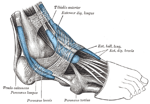
FIG. 1241 - The mucous sheaths of the tendons around the ankle. Lateral aspect. Arteries.—The femoral artery as it crosses the brim of the pelvis is readily felt; in its course down the thigh its pulsation becomes gradually more difficult of recognition. When the knee is flexed the pulsation of the popliteal artery can easily be detected in the popliteal fossa. 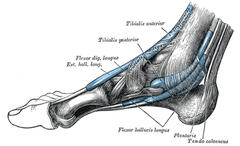
FIG. 1242 - The mucous sheaths of the tendons around the ankle. Medial aspect. On the lower part of the front of the tibia the anterior tibial artery becomes superficial and can be traced over the ankle into the dorsalis pedis; the latter can be followed to the proximal end of the first intermetatarsal space. The pulsation of the posterior tibial artery becomes evident near the lower end of the back of the tibia, and is easily detected behind the medial malleolus. Veins.—By compressing the proximal trunks, the venous arch on the dorsum of the foot, together with the great and small saphenous veins leading from it (see page 669), are rendered visible. Nerves.—The only nerve of the lower extremity which can be located by palpation is the common peroneal as it winds around the lateral side of the neck of the fibula.
Subscribe to the "News" RSS Feed 
TOP ۞